Oocyst culture: the lab variant
Culture conditions
The following technique works well for gametocysts from most terrestrial insects. Gametocysts collected from terrestrial hosts generally produce oocysts if they are protected from bacterial and fungal colonization in a moist environment at ambient temperature. Gametocysts collected from odonates and other aquatic hosts should be surface sterilized as described below and cultured in water individually using the field variant technique.
Surface sterilization
Stylocephalid gametocysts are protected by an outer epicyst and do not require surface sterilization. All other gametocysts should be surface sterilized by serial pipetting through the wells of a 3-welled dish. The first 2 wells are filled with water and the final well is filled with a 0.1% solution of Methyl Paraben in water.
Culture carriers
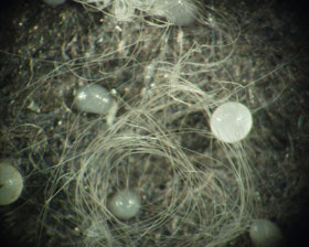
Gametocysts are much easier to handle, incubate, and store if they are placed on a carrier. In the lab variant technique, we use 6mm circles of black construction paper cut with a cork borer. Carrier dots are cut en mass and stored in a solution of 0.1% methyl paraben in water until needed. Presoaking the carrier dots leaches any excess dye and saturates the paper, improving capillary flow through the carrier dot. (NB: All brands of construction paper are not suitable. Some use heavy metal mordants in the dye process and these metals prevent gametocyst development. We find that the more expensive, archival papers work best.)
Loading carrier dots
Remove a carrier dot from the storage solution and place it on a piece of filter paper. The filter paper drains excess fluid from the carrier dot. As individual gametocysts are pipetted onto the carrier, excess transfer fluid is drawn through the carrier dot and absorbed by the filter paper. Once loaded, individual gametocysts should be arranged individually with a fine brush.
Incubation
Place carrier dots in the center of a 60-mm center-well organ cultuer dish (Falcon3037 or similar). We fill the outer well with a hydrating gel to maintain humidity for incubation. Individual culture dishes are covered and placed in a larger 100-mm glass petri dish to maintain humidity.
Making hydrating gel
The hydrating gel consists of cross-linked polyacrylamide gel (Soil Moist granules or similar) saturated with an aqueous solution of 0.1% Methyl Paraben (1 tablespoon of granules will suspend approximately 1 quart of solution). The resulting suspension is pureed in a blender to form a smooth hydrating paste. The hydrating gel stores for at least a year in screw top jars, so we make up several quarts at a time.